UC research team receives Brain Aneurysm Foundation grant
Researchers hoping to uncover a more accurate way to predict if an aneurysm will rupture

Researchers at the University of Cincinnati Department of Neurosurgery are hoping to uncover a more accurate way to predict if an aneurysm will rupture. Currently, providers look at the size of an aneurysm, but Charles J. Prestigiacomo, MD, and Mark Johnson, MD, are looking at the shape.
It is generally accepted that any aneurysm larger than 7 millimeters should be treated, when also taking into account other risk factors, such as patients who smoke, have hypertension or are female. Now, the team is hoping to define how to analyze an aneurysm’s shape to assess the risk of it rupturing.
“Subjectively, experts have described aneurysms as being irregular shaped or smooth shaped,” says Johnson, a fourth-year neurosurgery resident, “and the irregular aneurysms have been correlated in different population groups as more likely to rupture.”
The idea of studying an aneurysm’s shape is not new, but previously researchers have only looked at 2-dimensional shapes. UC neurosurgeons are looking at 3-D aneurysms. Under the direction of Prestigiacomo, the grant's principal investigator, Johnson and colleagues have developed a scoring system where a number can be assigned based on the morphological numerical.
“We’re using an interesting geometrical theorem,” Johnson said. “If we can validate that this works as an objective measure of that irregularity, then it is much easier to submit that integer to regression analysis to look at in a multi-variable model that could be used to help predict rupture risk, along with traditional risk factors.”
Essentially, the team hopes to turn this subjective way of using an aneurysm’s shape into an objective one.
That caught the attention of the Brain Aneurysm Foundation in spring 2022, when Johnson applied for a grant. The researchers secured the grant in July for the project, titled Morphological Analysis Through the Use of Fractal Dimension as a Predictor of Cerebral Aneurysm Behavior.

Mark Johnson, MD, presents his research. Photo provided.
Johnson has been combing through images of aneurysms in patients who have been treated at UC Health in the past seven years. He is hoping to transform between 500-700 of those images Into 3-D models.
“I make the 3-D model through a process called segmentation,” Johnson said. “I take the sourced images that were generated for clinical use and I put them into a software that turns the 2-D scans into a 3-D model. Then, I extract the aneurysm and do a shape analysis on it though a software program developed by former UC medical student Jimmy Castiligione.”
Initial pilot data of 30 patients suggests that Johnson and Prestigiacomo are on to something.
“There seems to be a strong correlation with prior rupture, more so than some of the other morphologic variables that have been tested,” Johnson said.
The pilot study only used data from computed tomography (CT) angiograms, and Johnson is hoping the correlation continues with different imaging modalities, like diagnostic angiograms and magnetic resonance angiograms.
“I want to make sure the measure you get with a DSA or MRA picture gives you a similar enough fractal dimension value that you can compare across the three imaging modalities,” he said.
An aneurysm’s growth will also be assessed to determine if it, too, could be used to predict risk of rupture. Additionally, Johnson will look at the fractal dimension of residual aneurysms to see if it is predictive of whether an aneurysm grows or resolves on its own.
Johnson aims to finish the study by September of 2023 and will be presenting the results of the trial at the Brain Aneurysm Foundation’s annual scientific meeting in Charleston, South Carolina.

Johnson and colleagues have received a $30,000 Brain Aneurysm Foundation grant to further their research. Photo provided.
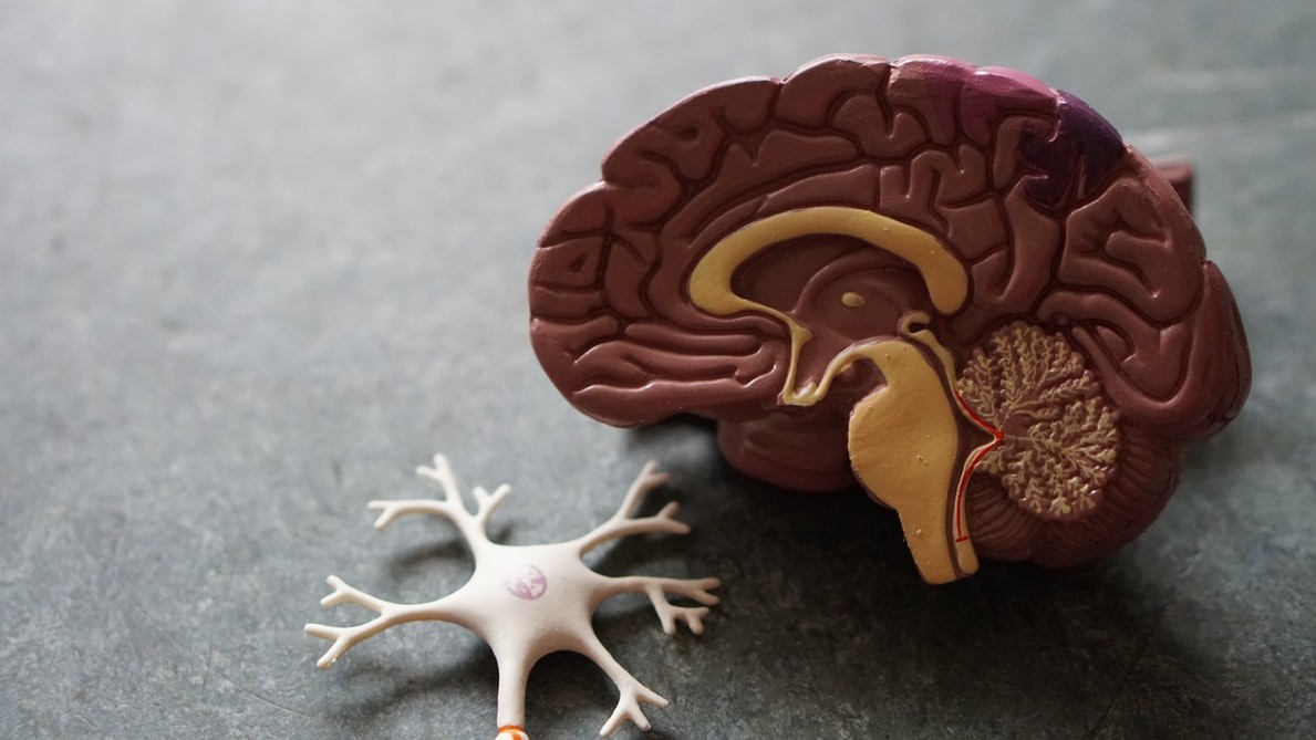
Featured photo at top of model of a brain. Photo/Robina Weermeijer/Unsplash.
Related Stories
Treating opioid use during pregnancy to take center stage during Addiction Center series
May 25, 2026
Join the University of Cincinnati on June 10 for a unique conversation on opioid use disorder during pregnancy, featuring landmark trial data and firsthand patient lived experience.
UC structural biologists are first in world to visualize key cell protein
May 22, 2026
University of Cincinnati College of Medicine structural biologists are the first in the world to visualize a key cell protein as part of recently published research in the journal Cell Reports.
6 ways starting a GLP-1 medication could affect your emotions
May 20, 2026
When patients first start taking a glucagon-like peptide-1 (GLP-1) medication, they probably expect to feel full. But they might not anticipate how it can influence their emotions. The medications act on the stomach and the brain, said Malti Vij, MD, a University of Cincinnati adjunct associate professor in the College of Medicine's Department of Internal Medicine and a diplomate of the American Board of Obesity Medicine.