‘Mini-brain’ shines light on concussions
Model shows how injuries create cascade of effects across cell, leading to neurodegenerative disease

A biomedical engineering professor at the University of Cincinnati is doggedly pursuing answers to one of medicine’s black boxes: concussions and other traumatic brain injuries.
Concussions are a common injury, responsible for as many as 3 million emergency room visits every year. Children playing sports or other recreation activities sustain nearly 4 million concussions every year, according to estimates by the Centers for Disease Control and Prevention.
UC College of Engineering and Applied Science Assistant Professor Volha “Olga” Liaudanskaya wants to know more about the milder, repetitive blows children in youth sports can sustain that can also lead to injury. In her lab at UC's Bioscience Center, she is studying how cells in the brain are affected by concussive forces — and how this trauma can lead to neurodegenerative diseases.
"We still know very little about what’s going on in the brain. And the injury can depend on the severity and location,” she said. “Brain trauma is so different from patient to patient, both the intensity of the injury and the patients themselves.”

UC researchers study the function of different types of brain cells using fluorescent dyes. Photo/Connor Boyle/UC Marketing + Brand
Using a ‘mini-brain’ to study concussions
Liaudanskaya and her team of student researchers are studying traumatic brain injury, or TBI, at the cellular level using novel models she created.
She calls them “mini-brains” and in her lab studies three brain cell types that regulate brain activity, including neurons. To that, she added two vascular cells, creating a complex “pentaculture” of five cell types that she can track simultaneously using living tissue.
“There was a big part missing, which was a vascular unit. For neurodegenerative diseases, the vascular system is a critical driver of inflammation, degeneration and proteinopathies like Alzheimer’s, Parkinson’s and chronic traumatic encephalopathy or CTE,” she said.
For people with a history of concussions or repeated blunt-force head trauma, understanding this vascular contribution is especially important.
We wanted to understand what mild injuries do to the brain. Is there a threshold? Is it an accumulation? Can we heal it?
Olga Liaudanskaya, UC College of Engineering and Applied Science
As most middle-schoolers learn, the mitochondria is “the powerhouse of the cell,” generating energy and regulating cellular processes.
“But it does so much more,” Liaudanskaya said. “Sex hormones, metabolism, epigenetic signatures — all are regulated by mitochondria. That’s why we wanted to look at them.”
Liaudanskaya is examining how mitochondria are affected and react to these concussive forces.
“The mitochondria plays a big role in neurodegeneration,” she said.
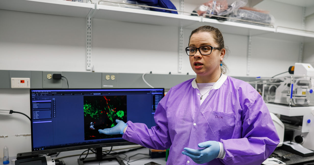
To study her mini-brains, she uses a specialized imaging tool called a confocal microscope that uses a laser and a pinhole aperture resulting in detailed — and extraordinary — 3D images that map the cells.
“We tagged mitochondria in neurons with a red fluorescent protein. You can see that here,” she said, pointing to a rotating 3D image on her computer screen. “We added green tags to the astrocytic mitochondria. You can see these large green cells. And white are the microglial mitochondria.”
Combined they create a detailed and colorful tapestry of the cellular function of the brain.
“We can track the mitochondria as it migrates in the cell. We can fluorescently tag mitochondria to see where it goes and what it does and what’s going on,” she said.

To study how cells respond to concussive blows, researchers first must deliver them using a custom device that mimics a blow to the head. In her biomedical engineering lab, UC Associate Professor Olga Liaudanskaya is studying how brain cells respond to head injuries that can cause concussions. Photo/Connor Boyle/UC Marketing + Brand
How blunt-force trauma affects brain cells
But first researchers have to subject the cells to concussive-like forces. They use mechanical devices that allow for consistent, measurable and replicable forces across samples, simulating blunt-force trauma that can cause concussions and traumatic brain injury.
Liaudanskaya’s most recent paper, published in the journal Frontiers in Cellular Neuroscience, examines the benefits of this computer modeling to study neurodevelopment and neurological disease. It’s one of the important ways her lab is tracking the cause and effect of blunt-force trauma through to the rise of neurodegenerative diseases. This concussion research helps explain how a single impact — or repeated mild head injuries — can create a cascade of cellular damage over time.
And she is not just looking at the traumatic brain injuries from car accidents or extreme collisions but the smaller, repetitive concussive forces that athletes, particularly children in youth sports, endure every season across sports.
“That was my dream because it’s not understood at all. There are no diagnostic markers out there of any kind,” she said.
Liaudanskaya said children are potentially exposed to less severe brain injuries more often than we know.
“So we wanted to understand what mild injuries do to the brain. Is there a threshold? Is it an accumulation? Can we heal it? What causes long-term neurodegeneration that is common in boxers and American football players,” she said, noting that these are key questions in TBI research.
Liaudanskaya found that in injured brain cells, certain proteins that normally stabilize neurons fail to return to their normal function. Fibers between nerve cells break down. There is chronic inflammation and metabolic dysfunction. And these are hallmarks of neurodegenerative disease.
Still, most people with concussions fully recover given enough time and the absence of any additional traumatic blows, she said.
“If you protect your brain for six months, you can recover. We see a pattern develop in the third month. Everything is stabilizing at the structural level, but you still have inflammation. If you extend it to six months and have another concussion, you start over,” she said.
Featured image at top: UC Associate Professor Olga Liaudanskaya is studying how brain cells respond to head injuries that lead to concussions. Photo/Connor Boyle/UC Marketing + Brand

Associate Professor Olga Liaudanskaya is studying the molecular processes of neurodegeneration caused by traumatic brain injury. By modeling concussions and mild traumatic brain injury with a lab-grown "mini-brain," University of Cincinnati engineers are uncovering how blunt-force impacts can trigger cellular cascades that may lead to long-term neurodegenerative disease. Photo/Connor Boyle/UC Marketing + Brand
FAQs: Repetitive brain injuries
Concussions and even milder traumatic brain injuries can trigger cellular dysfunction, leading to inflammation and disrupting communication between neurons. Over time, especially with repeated head impacts, cellular damage can contribute to a number of serious health issues. Traumatic brain injuries have been linked to conditions such as dementia, chronic traumatic encephalopathy, Alzheimer's disease, Parkinson's disease and mental health disorders such as depression.
This research is being conducted in the College of Engineering and Applied Science at the University of Cincinnati in Cincinnati, Ohio. UC biomedical engineers are collaborating with student researchers to advance concussion and traumatic brain injury science using innovative mini-brain models and advanced imaging tools.
A “mini-brain” is an in vitro model built from multiple types of human brain and vascular cells grown together in a lab. At the University of Cincinnati, researchers use this mini-brain model to study how concussive, blunt-force trauma affects brain cells at the cellular level.
Children who play sports can sustain injuries to their heads that do not result in diagnosed concussions. But University of Cincinnati researchers are using a novel mini-brain model to understand how less severe but repetitive injuries affect developing brains and whether there is a threshold or accumulation effect that increases the risk of long-term health issues.
The next groundbreaking discovery
UC is a powerhouse of discovery and impact as a Carnegie 1 research institution. From pioneering medical research to transformative engineering and social innovation, our faculty and students drive progress that reaches across the world.
Related Stories
How to prevent concussions in football? Better helmets
March 6, 2023
Football helmets made by four leading manufacturers showed vulnerabilities in tests designed to better understand player concussions, according to a study by the University of CIncinnati.
New Imaging Research & Development Center opens on UC’s medical campus
May 28, 2026
The University of Cincinnati’s new Imaging Research & Development Center on its Cincinnati medical campus brings UC, UC Health, Cincinnati Children’s, GE HealthCare and JobsOhio together to advance MRI research and accelerate imaging innovation.
UC lab prevents injuries with engineering
July 25, 2025
UC biomedical engineering Professor Eric Nauman and his students use biomedical engineering to prevent common injuries.