UC experts present neurology research at national conference
Spreading depolarization, robotic-guided thrombectomy insights highlight AAN abstracts

University of Cincinnati researchers will present abstracts at the 2026 American Academy of Neurology Annual Meeting April 18 to 22 in Chicago.
Study: Real-time diagnosis and treatment leads to fewer spreading depolarizations

Jed Hartings, PhD. Photo/University of Cincinnati.
Sometimes called a “brain tsunami,” a spreading depolarization (SD) is an event where brain cells lose their charge, become depolarized and are unable to send electrical signals to one another. SDs often occur following a traumatic brain injury and are associated with worse outcomes, but using this information to treat patients has not been previously tested.
A team led by UC’s Jed Hartings, PhD, Laura Ngwenya, MD, and Brandon Foreman, MD, are conducting the INDICT trial, the first Phase 2 trial to test the feasibility of treating patients based on diagnosing SDs in real time.
“We are trying to determine whether we can integrate information about spreading depolarizations into decision-making and treatment of severe traumatic brain injury,” said Hartings, professor and vice chair of research in the Department of Neurosurgery in UC’s College of Medicine. “In other words, can we use the information and methods developed in research on SDs and implement it for possible patient benefit in clinical care?”
Since 2023, researchers at UC, the University of California San Francisco and University of Pennsylvania have enrolled 31 patients in the trial.
“We have found that SDs can be diagnosed in real time, and that this information can be used to guide adjustments in clinical care,” Hartings said of early results from the trial. “Specifically, we have found that diagnosis and treatment result in a more than 50% reduction in the number of SDs that patients experience.”
As the trial moves forward, Hartings said an important step will be to determine whether identifying and treating SDs can result in long-term improved patient outcomes.
Hartings will present “Feasibility Trial of Spreading Depolarization Monitoring to Guide Management of Severe Traumatic Brain Injury: Initial Results.”
Researchers learn more about noninvasive detection of SDs

Brandon Foreman, MD. Photo/UC Health.
The best way to detect spreading depolarizations is to place an electrode strip within the brain, limiting most research to patients already requiring surgery for their injuries.
For patients who do not need surgery, clinical teams can monitor for SDs using a depth electrode placed at the bedside, known as clinical depth electrocorticography (dECOG).
UC researchers including first author Brandon Foreman, MD, conducted a retrospective review of 160 patients monitored for SDs after a traumatic brain injury.
“We found that the incidence of spreading depolarizations was much less with dECOG, likely related to the limited spatial sampling of this technique,” said Foreman, associate professor and an associate director of neurocritical care research in the Department of Neurology and Rehabilitation Medicine in UC’s College of Medicine and a UC Health neurocritical care physician. “However, the clinical significance of SD when they are detected is similar, suggesting that while you might pick up on SD less commonly, the use of bedside depth electrodes is better than not monitoring for SD at all.”
The team plans to expand its research to a larger number of patients treated at UC and other national and international datasets to better characterize SD detection using noninvasive methods.
“We are also participating in clinical studies in using advanced AI technologies to detect SD using standard scalp EEG, which is completely noninvasive,” Foreman said.
Foreman will present “The Incidence of Spreading Depolarizations in Severe Traumatic Brain Injury Detected by Depth Electrocorticography During Clinical Multimodal Neuromonitoring.”
Robotic-guided blood vessel navigation shows promise in preclinical study

Pablo Harker, MD. Photo/University of Cincinnati.
For some strokes, the best treatment is a thrombectomy, where doctors mechanically remove a blood clot in an artery to restore blood flow to the brain.
To accomplish this and some other neurovascular interventions, neurointerventionalists must precisely manipulate and navigate a guidewire through the blood vessels. Current guidewires are manipulated by the interventionalist from the groin or wrist with the tip of the wire within the blood vessels of the brain. Limited guidewire control can make procedures longer and increase the risk of complications.
Researchers from the Massachusetts Institute of Technology, Harvard Medical School and the University of Cincinnati, including first author Pablo Harker, MD, compared a novel magnetic robotic guidewire navigation system to conventional manual navigation. Using three anatomical models of increasing difficulty, four operators with varied levels of clinical experience performed a total of 72 procedures.
“We wanted to know if our novel magnetically steered, robotic system for navigating within the blood vessels of the brain improved the time it takes to access the target vessel,” said Harker, a fellow in UC’s Department of Neurology and Rehabilitation Medicine. “It reduces access times by a factor of 10 times and makes it so that there's a significantly smaller gap between experienced and junior interventionalists. This will result in shorter and safer procedures for patients requiring complex neurovascular interventions.”
Moving forward, the team plans to further develop the system and begin preclinical, “first-in-human” testing.
This research was financed by Magnendo Corp., a spinoff from the Department of Mechanical Engineering at MIT in collaboration with the Department of Neurosurgery at Massachusetts General Hospital/Harvard Medical School. Harker serves as a scientific advisor for Magnendo.
Harker will present “Closing the Experience Gap: Magnetic Robotic Navigation Enables Consistent Guidewire Performance Across Operators.”
Imaging data suggests link between glymphatic dysfunction, sleep apnea and reduced executive functioning
Cancer survivors commonly experience cancer-related cognitive impairment (CRCI), which can include sleep disorders such as obstructive sleep apnea that impair executive function.
Researchers including first author Sophie Kushman looked at the relationship between MRI markers of dysfunction of the glial lymphatic system, which removes waste material from the brain, and executive function test scores.
“Reduced clearance of toxic byproducts of normal brain metabolism and inflammation induced by cancer and its treatment could be contributing factors to CRCI and increased risk of neurodegenerative disease in cancer survivors,” said Kushman, a medical student at UC.
The team found several measures of executive function, including processing speed and sustained attention, declined with more enlarged perivascular spaces on MRI scans and increased sleep apnea severity.
“The high prevalence of sleep disturbances in CRCI and the relationship between executive function and EPVS burden shown here suggest treatments aimed at improving sleep disturbances, such as GLP-1 agonists, may regulate disrupted sleep-related cerebral spinal fluid flow and improve CRCI cognitive symptoms,” Kushman said.
The research was possible due to the clinical registry created at the University of Cincinnati Cancer Center’s cancer cognitive clinic, which helps track trends in this population.
“CRCI is complex and overlaps with risk factors associated with non-cancer cognitive impairment and neurodegenerative disease,” Kushman said. “Sleep is central to maintaining brain health. Understanding the relationship between sleep, glymphatic function and cancer is important for mitigating CRCI and neurodegenerative disease.”
Kushman will present “Imaging Proxies of Glymphatic Flow as a Mechanism for Executive Dysfunction in Cancer-related Cognitive Impairment.”
New program equips chief residents for unique leadership challenges

Jorge Patino, MD. Photo/University of Cincinnati.
Neurology chief residents play important administrative, academic and mentorship roles in shaping the learning environments at their institutions, but they are not given formal training to do so.
In collaboration with the American Academy of Neurology (AAN) and building on a successful program developed at New York University (NYU), researchers, including first author Jorge Patino, developed the Chief Resident Webinar Series, specifically designed to train future chief residents.
“Using their experience at NYU, we developed a one-year program comprising four asynchronous and four live sessions in which participants learn about feedback, graceful self-promotion, advocacy, recruitment and more,” said Patino, MD, a movement disorders fellow at UC’s James J. and Joan A. Gardner Family Center for Parkinson's Disease and Movement Disorders. “The program was very well received.”
The program is currently in its second year, with future, current and past chief residents welcome to participate and continue growing their leadership skills.
“The program has also been incorporated into the AAN Enhanced Resident Leadership Program, increasing its visibility and reach,” said Patino. “We hope that the CRWS will become a standard leadership training tool for all neurology residents in the United States.”
Patino will present “Leading the Future: A Neurology Webinar Series for Chief Resident Development” and “Writing for Publication” (W4P): Three-year Implementation and Evaluation of a Seminar-style Longitudinal Course in Scientific Writing for Academic Neurology.”
The next groundbreaking discovery
UC is a powerhouse of discovery and impact as a Carnegie 1 research institution. From pioneering medical research to transformative engineering and social innovation, our faculty and students drive progress that reaches across the world.
Other UC research being presented at AAN includes:
- Mohammadali Nahayati presenting "Safety and Efficacy of Teriflunomide on Clinical Course, and Laboratory Findings in Patients With HTLV‑1‑associated Myelopathy/Tropical Spastic Paraparesis."
- Vincent Martin presenting "Evaluation of Unmet Needs and Quality-of-Care Indicators among Patients with Migraine in the United States: 2021–2022" and "Treatment with Fremanezumab Attenuates the Association Between Weather and Headache in Participants with Episodic Migraine: A Post Hoc Analysis of the HALO-EM and HALO-LTS Studies"
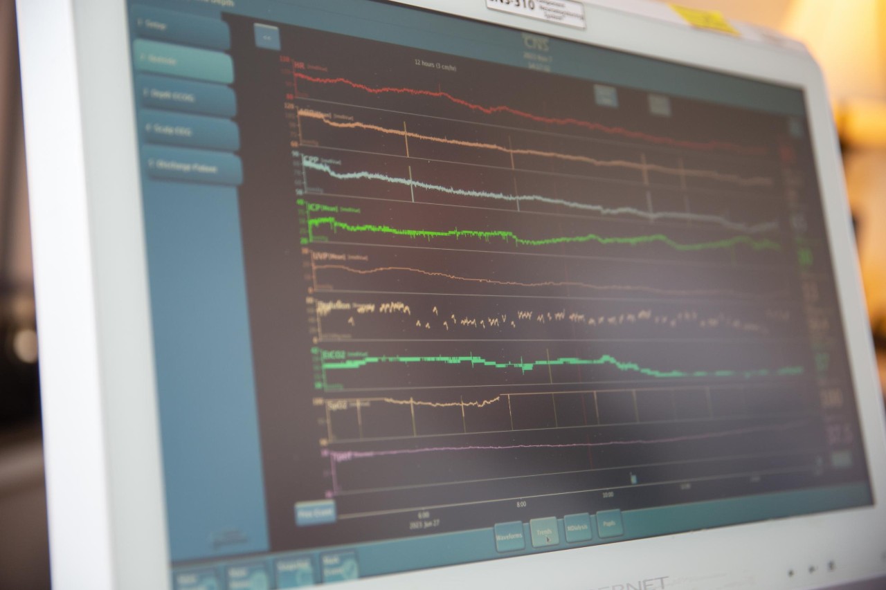
Featured photo at top of neuromonitoring after a traumatic brain injury. Photo/Andrew Higley/UC Marketing + Brand.
Related Stories
Searching for a cure for deadly brain tumors
March 24, 2022
The University of Cincinnati is enrolling patients for a new clinical trial testing a two-pronged immunotherapy approach to treat glioblastomas, deadly brain tumors.
A potential new treatment for brain tumors
September 23, 2022
The University of Cincinnati's Pankaj Desai, PhD, has received a $1.19 million grant from the National Institutes of Health/National Institute of Neurological Disorders and Stroke to continue research into the use of a drug called letrozole to treat glioblastomas, the most deadly form of brain tumors.
Collaborative University of Cincinnati Cancer Center team opens Phase 2 brain tumor trial
March 26, 2024
A multidisciplinary team of University of Cincinnati Cancer Center researchers have opened a Phase 2 clinical trial to test a new combination treatment for glioblastomas, the most deadly form of brain tumors.