UC Surgeons Report New Treatment for Often-Fatal Injury

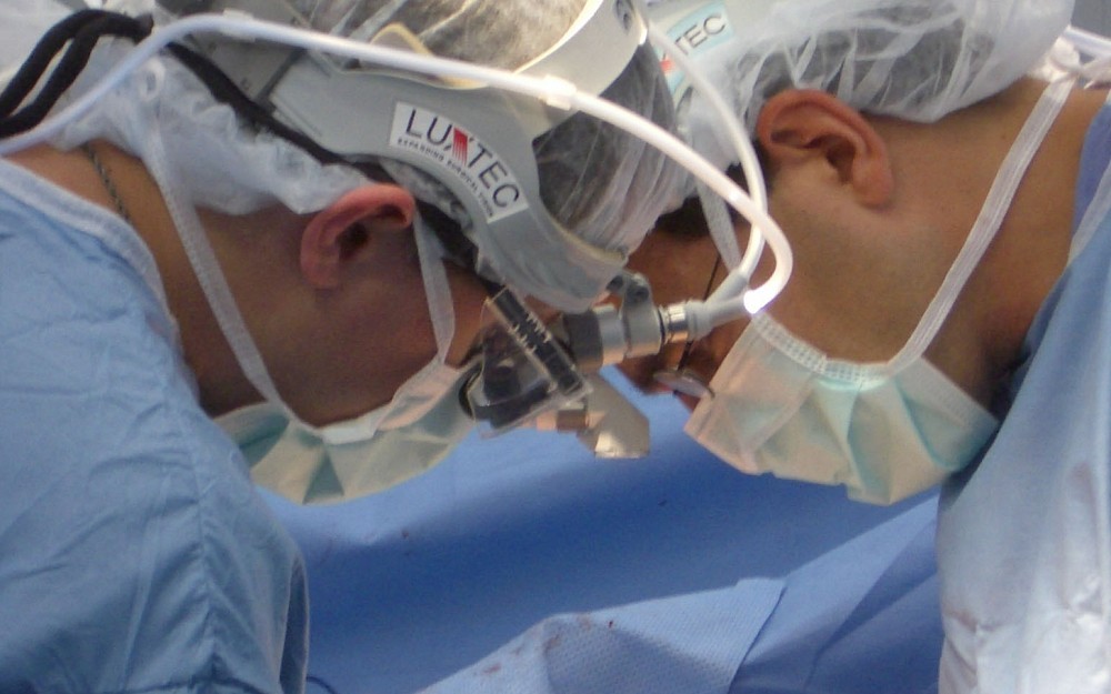
CINCINNATIUniversity of Cincinnati (UC) surgeons have developed a new, minimally invasive method for repairing a common and deadly form of aortic injuryan advance that could help reduce the number of deaths caused by auto accidents and major falls.
The potentially life-saving technique is reported by Joseph Giglia, MD, interim director of UCs division of vascular surgery, and his team in the March edition of Annals of Vascular Surgery.
Dr. Giglia estimates that 80 to 90 percent of the patients who suffer this injuryknown as blunt thoracic aortic traumadie before ever reaching a hospital.
The aorta is the main artery that carries oxygenated blood from the heart to be distributed throughout the body.
A severe blunt thoracic accident, usually a non-penetrating deceleration injury, can jerk the aorta forward and back again, causing it to crack.
If all three layers of the aorta rupture immediately, the patient will die almost instantly from blood loss. If the outer layer remains intact, however, a weakened area called a pseudoaneurysm can form, which often bursts later.
Surgeons must repair the damaged aorta quickly before it does rupture, killing the patient.
The Food and Drug Administration has yet to approve a device for treatment of this type of injury, says Dr. Giglia, and custom-made devices are usually large and difficult to use in an emergency setting.
Dr. Giglias team found that by placing a standard surgical stenta tube-shaped metallic support structurein the aorta and lining it with a series of small synthetic grafts (endografts) they could effectively protect the damaged area without stopping blood flow to the rest of the body.
The endografts are regularly used in vascular surgery to treat aneurysms in the abdomen, Dr. Giglia explains, but they are too short to fully bridge the damaged area in the chest.
Used alone, extender cuffs are hard to align and often slip into the defect to cause further complications, says Dr. Giglia. Using a fixed stent provides an easy-to-see scaffolding that can guide the surgeon to more accurately secure the extender cuffs and create a strong seal.
Once in place, the dual lining supports the weakened vessel walls and allows blood to continue flowing through the aorta without applying pressure to damaged area.
Our method gives emergency surgeons a reliable way to repair difficult injuries with readily accessible materials, Dr. Giglia says. More people can be treatedand potentially savedwith these minimally invasive techniques.
-30-
Tags
Related Stories
Cancer Center plays key role in lab research, clinical trials for new pancreatic cancer drug
September 1, 2026

WLWT and WCPO highlighted the contributions of University of Cincinnati Cancer Center researchers that helped lead to the recent FDA approval of daraxonrasib, a new treatment for pancreatic cancer.
Cancer Center physician receives $150,000 New Investigator Research Grant to study treatment-resistant leukemia
September 1, 2026
The University of Cincinnati Cancer Center’s Sarah Skuli, MD, PhD, received a two‑year, $150,000 Leukemia Research Foundation New Investigator Grant to study TP53‑mutated leukemia, a treatment‑resistant subtype with very poor outcomes.
Heavy social media use linked to depression
August 31, 2026

U.S. News and World Report, Yahoo! News, MSN, USA Today and other national media highlight a new study by the University of Cincinnati and Northwestern University that found a correlation between heavy social media use and depression in adults.