Combined Imaging Approach May Provide Better Identification of Difficult-to-Diagnose Brain Clots

CINCINNATIUniversity of Cincinnati (UC) neuroradiologists believe a brain imaging approach that combines standard magnetic resonance imaging (MRI) scans with specialized contrast-enhanced techniques could lead to more effective diagnoses in patients with difficult-to-detect blood clots in veins of the brain.
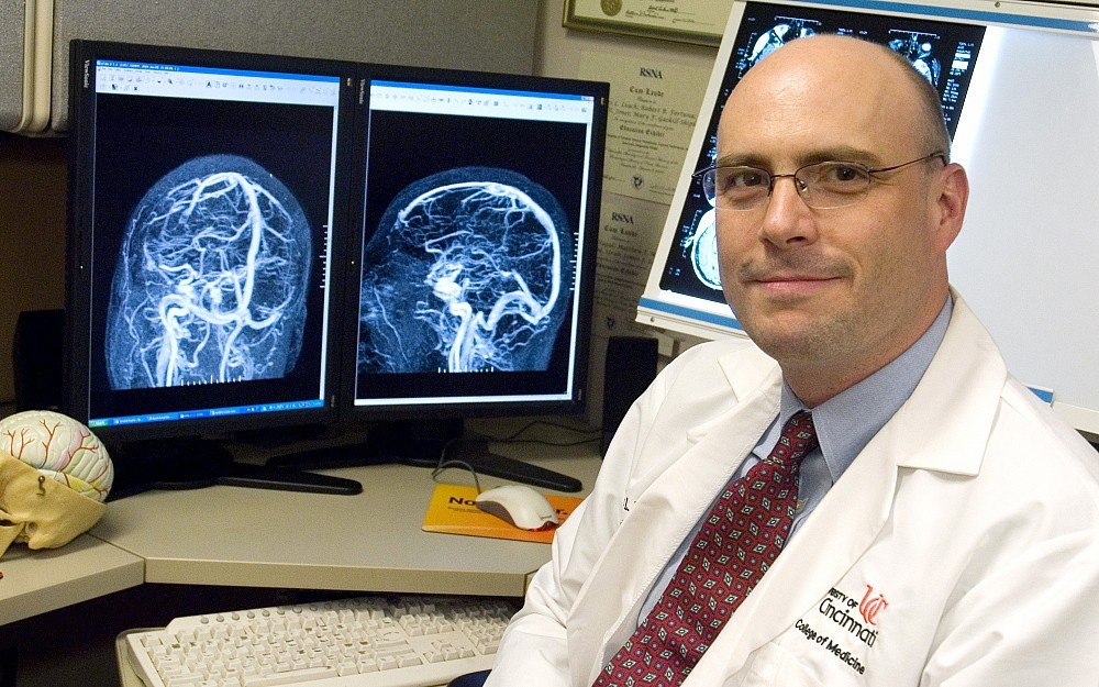
James Leach, MD, reports these findings in the April issue of the American Journal of Neuroradiology. This is the first study to correlate the clinical importance of data gleaned from standard MRI scans and detailed contrast-enhanced imaging techniques in patients with chronic thrombosis (blood clots) in veins of the brain.
Detailed contrast-enhanced techniques produce more defined distinctions between abnormal and normal veins in the membrane around the brain, explains Leach, a neuroradiologist and associate professor at UC and principal investigator of the study. Evaluating patients using a combination of imaging tools could give us a better understanding of the disease process.
Researchers say these specialized techniquesknown as contrast-enhanced magnetic resonance venographyproduce better defined pictures of difficult-to-detect abnormal flow areas within vessels of the brain. These areas may be important warning signs of blocked blood flow that require medical intervention. The enhanced imaging tools can also help identify areas where flow has been partially reestablished after a vessel blockage has occurred.
Leach and his team used these contrast-enhanced techniques, in combination with standard MRI scans, to evaluate a small subgroup of patients who have clinical and imaging features consistent with what is called partially recanalized chronic dural sinus thrombosis. This is a condition in the membrane surrounding the brain where blood flow has been partially reestablished in a previously blocked vessel.
Research has shown that cerebral veins partially or fully blocked by a blood clotwith or without treatmentcan open spontaneously, open partially or remain blocked. These blockages can stop blood flow to the brain and increase the patients risk for stroke and other neurological episodes such as chronic headache.
In cases where the patient has an ongoing partial or full blockage, Leach explains, the blood clot organizes into a more solid lesion, forms connective scar tissue and can develop small vessels in an attempt to restore blood flow. This was very difficult to diagnose correctly using previous techniques."
We dont fully understand why this happens, but being able to recognize the imaging characteristics of the condition earlier may help us explain patient symptomslike persistent headachethat otherwise cannot be explained, he adds.
With additional research, Leach believes, this combined imaging approach may one day help physicians formulate better treatment regimens and possibly prevent more serious medical complications for certain patients.
If we can more easily characterize the patients problem as chronic instead of acute, it could alter the way we prescribe anticoagulation (clot-busting) drugs to treat the symptoms, says Leach. Further research is needed to see if this can effectively impact patient therapy clinically.
Collaborators in this study include Michael Wolujewicz, MD, and William Strub, MD, who are both affiliated with the UC College of Medicine and University Hospital.
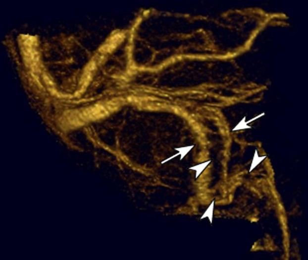
This image from a high detail contrast-enhanced MR venogram shows areas of restored blood flow (arrows) and areas of persistent blood clot (arrowheads).
Related Stories
Driven by curiosity, guided by care
May 14, 2026
Max Wilson, a University of Cincinnati College of Allied Health Sciences health sciences major on the pre-physician assistant track, found his path expanding beyond the classroom and into hands-on research focused on human performance and patient care.
Is a colonoscopy painful?
May 13, 2026
The University of Cincinnati's Susan Kais, MD, assistant professor of clinical medicine in the Division of Gastroenterology and Hepatology in the College of Medicine and UC Health gastroenterologist, recently appeared on the ARC Cincinnati morning program on Local 12/WKRC-TV to answer common questions from viewers about colonoscopies and to dispel myths.
University of Cincinnati graduate programs rise in national rankings across high-demand fields
May 13, 2026
University of Cincinnati graduate programs climbed in the latest U.S. News & World Report rankings, led by strong gains in workforce-focused fields including public health, clinical psychology and business.