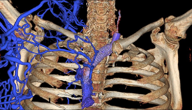
New Thoracic Imaging Approach Can Pinpoint Underlying Venous Problems

University of Cincinnati (UC) radiologists have developed a new technique for capturing images of chest veins that eases diagnosis of venous diseases.
Multi-detector computed tomography (CT) scanners are traditionally used to create three-dimensional images of arteries, the vessels which carry oxygen-rich blood away from the heart and distribute blood throughout the body. Veins, smaller vessels that return blood to the heart, are more difficult to accurately image.
Developed by Cristopher Meyer,
We found that the rapid-imaging scanners were almost too fast for venous studies, explains Vagal, a UC assistant professor and radiologist at
Venous disease is rare and can be difficult to pinpoint, she adds. This new protocol uses the same imaging equipment in a novel way that allows us to acquire better venous images and make good clinical decisions.
Vagal presented guidelines for this thoracic imaging protocol at the North American Society of Cardiovascular Imagings 35th Annual Meeting and Scientific Sessions in
For this new imaging technique, the CT technologist prepares two syringes of contrast: The first includes 140 cubic centimeters (CC) of undiluted contrast; the second contains a diluted mixture of 100 CC of contrast and 10 CC of saline solution.
The key to getting accurate clinical images of the veins is in the timing, Vagal says.
Both syringes are given consecutively at a rate of four CC per second, with a 60-second delay between the final injection and initiation of the CT scan.
Previously, there was so much dense contrast in the veins that all you could see on the CT scan were streaks that didnt tell you anything about possible venous disease, explains Vagal. Delaying the scan gave us enough time for both the arteries and the veins to be opacified, which resulted in the crisp images that allowed us to make better clinical determinations.
Vagal is affiliated with the Neuroscience Institute at UC and University Hospital, a center of excellence that focuses on the main diseases of the brain and nerves such as stroke, brain tumors, brain trauma, Parkinsons and Alzheimers disease, epilepsy, ALS and multiple sclerosis.

Achala Vagal, MD, is an associate professor of radiology and neuroradiologist with UC Health.
Tags
Related Stories
On-site training in Speech & Hearing Clinic sets UC’s programs apart
August 27, 2026
At the University of Cincinnati’s Speech & Hearing Clinic, Doctor of Audiology and Speech-Language Pathology graduate students gain real-world experience while providing free speech and hearing services to the community.
Funding expands UC research into weight gain side effects of bipolar medication
August 26, 2026
University of Cincinnati researchers have been approved for funding by the Patient-Centered Outcomes Research Institute (PCORI) to expand the implementation of healthy lifestyle interventions and metformin prescription to help manage weight gain in overweight or obese youth taking second-generation antipsychotic medications.
One in four NFL players had CTE at death
August 25, 2026

Forbes cites research on concussions and football helmets by the University of Cincinnati in a report that found one in four NFL players had brain damage at death.