Earlier Detection of Breast Tumors Is Goal of UC Study

CINCINNATIA new study at UC Health University Hospital involving technology that is not being used anyplace else in Cincinnati may help specialists find breast tumors in the earliest stages and biopsy them at the time of detection.
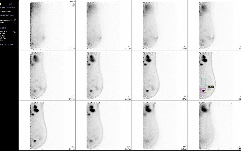
Amy Argus, MD, UC Health radiologist and investigator on the study, says the new technologypositron emission mammography (PEM)uses radioactive glucose to help image tumors as small as 2 millimeters in size.
"We inject glucose into the patients bloodstream intravenously, she says. "Tumors have a higher metabolism and tend to take up more of the glucose and trap it. The imaging is similar to a standard mammogram, but the panels are able to make the radioactive glucose visible.
Argus says the high-resolution images resulting from PEM could help physicians determine how to move forward with diagnosis and treatment.
"PEM has been around for a while as a diagnostic tool, she says. "Other studies have compared it to breast MRI, which is often used to find tumors. These studies suggest that PEM is as sensitive and/or more specific, meaning that it gives us less false positives, preventing us from taking biopsies on non-cancerous tissue.
Researchers in this study will couple an investigational biopsy method with PEM to allow physicians to take a biopsy using PEM images.
"This is important because sometimes were only able to see abnormalities with PEM, Argus says. "If an abnormality cannot be seen any other way, it is difficult to be sure that a biopsy is done in the correct location. In being able to take the biopsy using PEM, were able to get a more accurate sample.
Argus says this technology could be extremely promising for patients who have been recently diagnosed with breast cancer.
"PEM could help us find additional tumors or cancer cells that may have been missed by mammographyareas with additional disease, she says. "We believe we will be able to make a diagnosis and deliver treatment faster by doing imaging and biopsies at the same time.
"Prior breast MRI research shows that 15 to 30 percent of patients have more disease that does not show during a standard mammography. This has the potential to help physicians find the disease and treat it in the quickest, most efficient way possible.
Naviscan, the makers of the PEM Flex Solo II scanner, provided the equipment for this study. Argus receives no honoraria from Naviscan and cites no conflict of interest.

Amy Argus, MD, UC Health radiologist
Tags
Related Stories
Cancer Center plays key role in lab research, clinical trials for new pancreatic cancer drug
September 1, 2026

WLWT and WCPO highlighted the contributions of University of Cincinnati Cancer Center researchers that helped lead to the recent FDA approval of daraxonrasib, a new treatment for pancreatic cancer.
Cancer Center physician receives $150,000 New Investigator Research Grant to study treatment-resistant leukemia
September 1, 2026
The University of Cincinnati Cancer Center’s Sarah Skuli, MD, PhD, received a two‑year, $150,000 Leukemia Research Foundation New Investigator Grant to study TP53‑mutated leukemia, a treatment‑resistant subtype with very poor outcomes.
Heavy social media use linked to depression
August 31, 2026

U.S. News and World Report, Yahoo! News, MSN, USA Today and other national media highlight a new study by the University of Cincinnati and Northwestern University that found a correlation between heavy social media use and depression in adults.