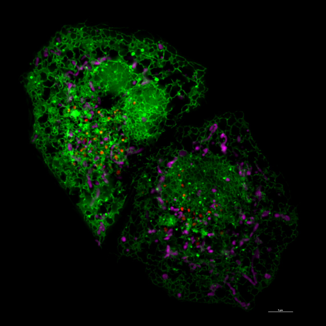
55KRC: UC research advances view at the subcellular level

University of Cincinnati cancer biologists have developed a new piece of technology and a new imaging technique that will help researchers glean more detailed data points and see cells in more precise detail when studying the development of cancer and neurodegenerative diseases.
Jiajie Diao, PhD, associate professor in the Department of Cancer Biology in UC’s College of Medicine, told 55KRC's Simply Medicine that a new probe his team developed helps give more details about the cellular environment, such as pH level.
The probe, combined with a new imaging technique that helps measure the shape, distance and location of tiny parts within cells called organelles. Seeing how the organelles interact can provide more information to how these interactions lead to the development of diseases, he said.
"We are actually working with the most advanced image technique to develop new analysis methods, trying to understand and trying to review subtle changes, changes you couldn’t even notice by your eyes," Diao said. "We’re trying to catch diseases at a very early, early stage."
Listen to the Simply Medicine segment. (Note: Segment begins around 23:40 mark.)
Featured photo at top of super resolution image of cell courtesy of Jiajie Diao.
Related Stories
Get to know CCM’s newest faculty and staff members
July 29, 2026
UC’s College-Conservatory of Music will welcome a variety of new faculty and staff members to its roster of distinguished performing and media arts experts, researchers and educators this fall.
UC co-op helps build career pathways at AtriCure
July 28, 2026
AtriCure, a global medical device company in Mason, Ohio, is UC’s largest biomedical engineering co-op employer, turning University of Cincinnati co-op students into full-time innovators.
CCM welcomes new musical theatre faculty member Avery Bargassé
July 28, 2026
UC College-Conservatory of Music Dean Pete Jutras has announced the appointment of Avery Bargassé as CCM's new Assistant Professor Educator of Musical Theatre Voice. His faculty appointment officially begins on Aug. 15, 2026. Bargassé is a versatile vocalist, recording artist and voice teacher whose career spans television, popular music and classical singing. He approaches teaching from a whole-person perspective, helping students develop healthy, flexible technique that supports expressive communication across styles.