55KRC: UC research advances view at the subcellular level

University of Cincinnati cancer biologists have developed a new piece of technology and a new imaging technique that will help researchers glean more detailed data points and see cells in more precise detail when studying the development of cancer and neurodegenerative diseases.
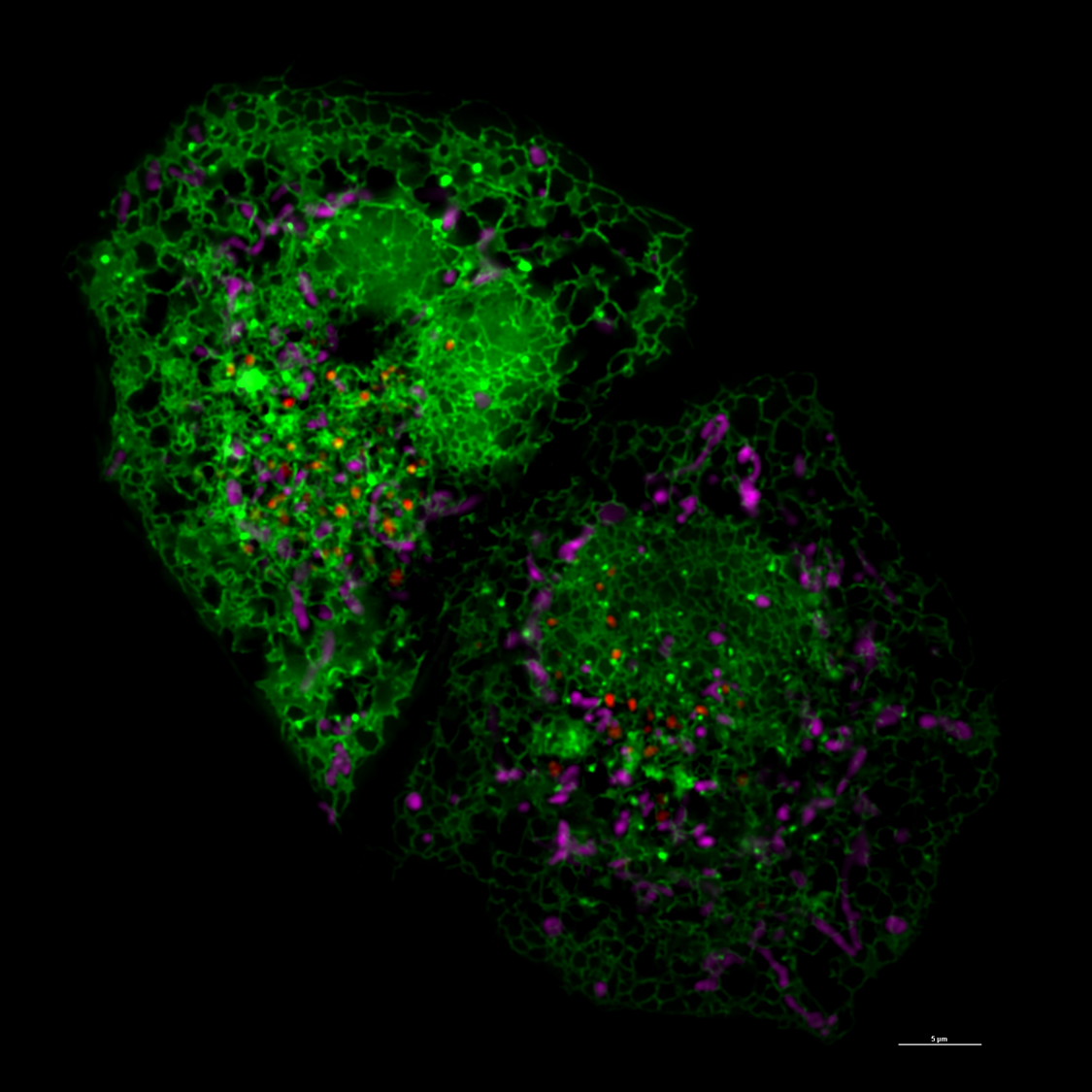
Jiajie Diao, PhD, associate professor in the Department of Cancer Biology in UC’s College of Medicine, told 55KRC's Simply Medicine that a new probe his team developed helps give more details about the cellular environment, such as pH level.
The probe, combined with a new imaging technique that helps measure the shape, distance and location of tiny parts within cells called organelles. Seeing how the organelles interact can provide more information to how these interactions lead to the development of diseases, he said.
"We are actually working with the most advanced image technique to develop new analysis methods, trying to understand and trying to review subtle changes, changes you couldn’t even notice by your eyes," Diao said. "We’re trying to catch diseases at a very early, early stage."
Listen to the Simply Medicine segment. (Note: Segment begins around 23:40 mark.)
Featured photo at top of super resolution image of cell courtesy of Jiajie Diao.
Related Stories
UC doctoral student shares his journey with Spectrum News
July 27, 2026
UC doctoral student Amota Ataneka spoke with Spectrum News about his educational journey. Ataneka, a native of the Pacific island nation of Kiribati, is one of 35 emerging scholars across the United States to win the National Academy of Education/Spencer Dissertation Fellowship.
How scammers can persuade you with AI
July 27, 2026
Spectrum News recently dove into how cloning voices with artificial intelligence is making it easier to scam people over the phone. The news outlet spoke with Kimberly Hyun, assistant professor of marketing at UC’s Lindner College of Business, to examine why AI is making it so much easier to trick people.
Decade of discovery
July 27, 2026
Every summer, UHP Discover has connected undergraduate students in the University Honors Program (UHP) to a unique array of opportunities in original interdisciplinary research. This year, the program celebrates its 10th anniversary.