Study finds distinct patterns of preexisting brain health characteristics in stroke patients
UC stroke researchers present at international conference

University of Cincinnati researchers are presenting abstracts at the European Stroke Organization Conference (ESOC) 2023, May 24-26 in Munich, Germany, including the results of the first large-scale assessment of radiological brain health in stroke patients in a population.
Extensive research has helped pinpoint risk factors for initial stroke, but there is limited understanding about what the brains of stroke patients look like on a population level, according to UC’s Achala Vagal, MD, professor of neuroradiology.
“Imaging can be an objective manifestation of the presence and severity of clinical factors such as diabetes, hypertension, high cholesterol and kidney failure,” she said. “However, the majority of the large epidemiological studies of brain health have been performed in stroke-free subjects.”
Vagal was a co-principal investigator on the Assessing Population-based Radiological brain health in Stroke Epidemiology (APRISE) study that gained new information from neuroimaging results of stroke patients.

Achala Vagal, MD. Photo/Ravenna Rutledge/UC Marketing + Brand.
The research team analyzed all available clinical imaging data from nearly 3,500 patients who had a stroke in the Greater Cincinnati/Northern Kentucky region in 2015, assessing the imaging for signs of small vessel disease in the brain in the form of previous injury, microbleeds, white matter disease (wearing away of tissue) or brain atrophy, among other observations.
Vagal said the team identified three distinct clusters of observable imaging characteristics that were each associated with a specific set of clinical variables.
“This can help us understand the biology of preexisting brain health in stroke patients and help guide future interventions,” she said. “We expected all the imaging parameters of brain health due to small vessel disease to be closely clustered, but we found a lack of clustering of microbleeds with white matter disease.”
With the knowledge gained from the study, Vagal said the team is now using the brain health imaging data to build a prediction model of recurrent stroke.
“Such large-scale characterization of preexisting brain health is helpful to identify novel observable characteristics which can guide further studies,” she said.
Vagal presented the oral abstract “Radiological Phenotypes of Brain Health in a Stroke Population: Primary Results of the Assessing Population-Based Radiological Brain Health in Stroke Epidemiology (APRISE) Study'' during the Imaging scientific communication session Thursday, May 25.
Other abstract co-authors include UC’s Heidi Sucharew, Vivek Khandwala, Lily Wang, Rebecca Cornelius, Mary Gaskill-Shipley, Thomas Tomsick, Shantala Gangatirkar, Brady Williamson, Thomas Maloney, Mary Haverbusch, Janice Carrozzella, Kathy Alwell, David Robinson, Robert Stanton and co-principal investigators Pooja Khatri, MD, and Brett Kissela, MD; Paul Horn of Cincinnati Children’s Hospital Medical Center; David Wang of the I-MED Radiology Network in Melbourne, Australia; and Dawn O. Kleindorfer of the University of Michigan.
Other presentations from UC researchers include:
- Felipe Ayala, “Collateral Grade as a Mediator of the Effect of Post-Endovascular Blood Pressure Goals on Outcomes: A Pre-specified Exploratory Analysis of the BEST-2 Randomized Trial,” during the Late Breaking Moderated poster session Thursday, May 25.
- Joseph Broderick, “RFVIIA for Acute Hemorrhagic Stroke Administered at Earliest Time (FASTEST) Trial,” during the Ongoing Trials poster session Thursday, May 25.
- Destiny Hooper, “Diffusion-Weighted Image Positivity Predicts Three-Year Risk of Stroke or Death After Transient Ischemic Attack in Biracial Population,” Wednesday, May 24.
- Hooper, “Elevated Leukocyte Counts Are Associated With Worse Functional Outcomes in Patients with Large Vessel Occlusion,” during the Prognosis and Outcome After Stroke poster session May 25.
- Pooja Khatri, “Ischemic Stroke with Minor Disabling Deficit,” during the Improving Outcomes of Acute Reperfusion Therapies for Acute Ischemic Stroke scientific session May 26.
Next Lives Here
The University of Cincinnati is leading public urban universities into a new era of innovation and impact. Our faculty, staff and students are saving lives, changing outcomes and bending the future in our city's direction. Next Lives Here.
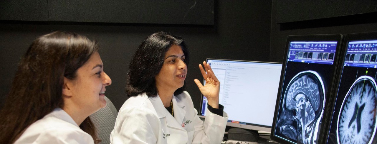
Featured photo at top of Pooja Khatri, left, and Achala Vagal, right, reviewing brain images. Photo/Ravenna Rutledge/UC Marketing + Brand.
Related Stories
The future, decoded. UC scholars reveal what’s next
May 20, 2026
The University of Cincinnati’s NEXT Innovation Scholars presented Gen Z-fueled insights on possible future trends at the Futures Forum 2026. Here’s what they see ahead.
CCM welcomes new film and media scoring faculty member J.R. Paredes
May 20, 2026
UC College-Conservatory of Music Dean Pete Jutras has announced the appointment of J.R. Paredes as CCM's new Assistant Professor of Film and Media Scoring. His faculty appointment officially begins on Aug. 15, 2026. Paredes is a composer, music producer and audio post-production specialist whose work spans film, television and commercial music. His credits include original scores for feature films and series distributed on platforms such as Apple TV+ and Prime Video, as well as extensive work in sound design and mixing for film and media.
6 ways starting a GLP-1 medication could affect your emotions
May 20, 2026
When patients first start taking a glucagon-like peptide-1 (GLP-1) medication, they probably expect to feel full. But they might not anticipate how it can influence their emotions. The medications act on the stomach and the brain, said Malti Vij, MD, a University of Cincinnati adjunct associate professor in the College of Medicine's Department of Internal Medicine and a diplomate of the American Board of Obesity Medicine.