Arming the body’s immune cells
UC scientists have uncovered a mechanism that could lead to new immunotherapies head and neck cancer

Researchers at UC have discovered a previously unknown mechanism that could explain the reason behind decreased immune function in cancer patients and could be a new therapeutic target for immunotherapy for those with head and neck cancers.
The authors share these findings in an article published in the journal Frontiers in Pharmacology.
Immunotherapy is a type of cancer treatment that boosts the body's natural defenses to fight cancer.

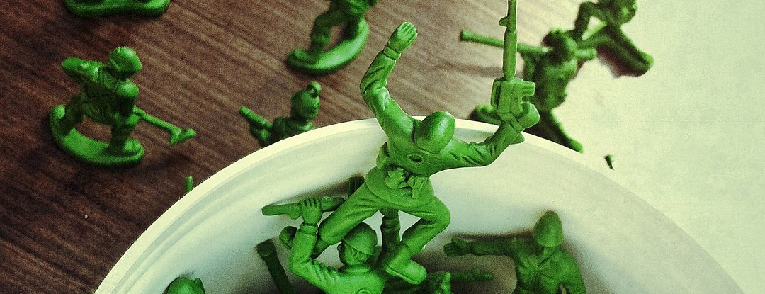
Ameet Chimote, first author on the study, says cytotoxic T-cells are like the soldiers of our immune system and are our body's first line of defense against cancerous tumors. Photo/Colleen Kelley/UC Creative Services
Led by Laura Conforti, professor in the Department of Nephrology and Hypertension at the UC College of Medicine, the team discovered that a reduced interaction between a molecule called calmodulin and an ion channel (KCa3.1) in the immune cells of cancer patients plays an important role in the reduced function of these cells. The team performed experiments on white blood cells called cytotoxic T-cells taken from the blood of patients with head and neck cancer.
“Cytotoxic T-cells are like the soldiers of our immune system and are our body's first line of defense against cancerous tumors,” says first author Ameet Chimote, research scientist in Conforti’s laboratory. “Just like how soldiers on the frontlines penetrate the enemy's defense and launch a massive attack, these cytotoxic T-cells are expected to penetrate the solid tumors by migrating within the tumor mass and then secreting chemicals called cytokines to kill these tumor cells. Sadly, for some reason, these cells do not function properly in patients with cancer, and they do not penetrate the tumors and attack the tumor cells, causing the cancerous tumors to grow uncontrollably.”

Corresponding author Laura Conforti, professor in the Department of Nephrology and Hypertension, and Ameet Chimote, research scientist, in Conforti's lab. Photo/Ameet Chimote.
“Identifying the mechanism of this underlying dysfunction can help us identify molecules that we can target with drugs and ultimately restore the ability of these cells to enter and kill the tumors,” says Conforti. She says that molecules, known as ion channels, are present in the T-cell membranes and are essential for T-cell function.
"In this study, we were able to show that the function of these channels in T-cells from cancer patients is decreased which results in a decreased T-cell accumulation in solid tumors,” Conforti says.
These types of channels require a signaling molecule called calmodulin to bind to them in order to function to their full capacity; this is needed even more in cancer T-cells.
Using several intricate microscopy imaging techniques on T-cells isolated from the blood of cancer patients, the team found out that, as compared to T-cells from healthy individuals, the cancer T-cells have fewer calmodulin molecules in their membranes.

“This would mean that there is less calmodulin binding to the channels in the T-cells from cancer patients,” Conforti says. “As previously stated, the channels do not function if the calmodulin does not bind to them. Thus, the decreased calmodulin binding in T-cells from cancer patients results in decreased function and leads to reduced tumor infiltration and killing of the cancer cells.”
“So back to our soldiers: If they were present at a battlefield, but none of them have any weaponry, this will hinder their ability to infiltrate the enemy lines. Now, if we arm each soldier adequately, we would boost their function. They can do their job. Similarly, we observed that if we increase the function of these channels by drugs that enhance their activity, the cancer patients' T-cells can penetrate the tumors better and also produce increased cytokines which can kill tumor cells,” Chimote says. “These are exciting findings that could lead to additional treatments for patients with cancer.”
“These findings strengthen the therapeutic potentials of [these] activators, which could restore cytotoxic T-cell functionality and can ultimately lead to additional immunotherapeutic options for patients with cancer,” Conforti adds.
Featured photo/Maclapessoa. Ameet Chimote, first author on the study, compared cytotoxic T-cells to soldiers or Army men.
Next Lives Here
The University of Cincinnati is classified as a Research 1 institution by the Carnegie Commission and is ranked in the National Science Foundation's Top-35 public research universities. UC's graduate students and faculty investigate problems and innovate solutions with real-world impact. Next Lives Here.
This work was funded by grant support from the NIH (grant R01CA95286, T32CA117846T) and a Clinical and Translational Science Award (CTSA) — awarded KL2 Mentored grant and a grant from CCTST (1UL1TR001425-01). Researchers cite no conflict of interest.
Related Stories
UC Foundation Board of Trustees announces seven new members
June 17, 2026
The University of Cincinnati Foundation is proud to announce that seven dedicated community members are joining its board of trustees. Ray Brooks, CEAS ’83; Chris Carper, A&S ’93; Tim Giglio, Bus ’84; Nandita Jena, A&S '03, Bus '03 and Chris Lewis, MD, Med ’00 will begin their term in October 2026. In addition to this esteemed group, two new student trustees, Case Trokhan, Med ’26 and Joseph Verry, Med ’30 are joining the board.
UC alumni set ‘gold standard’ for scrap metal business
June 17, 2026
University of Cincinnati graduates Finn and Bowen Alexy have found tremendous business success in their lower 20s as leaders at brazing company Alexy Metals and co-founders of venture firm BowFin Capital.
GBBN presents Creative Impact Award during inaugural DAAPworks 2026 ceremony
June 17, 2026
GBBN served as industry jury partner and presented Creative Impact Awards during DAAPworks 2026, recognizing exemplary architecture student projects at UC during the inaugural DAAPworks ceremony led by new DAAP Dean Stephanie Pilat.