Advancing our view at the subcellular level
UC researchers develop new probe, imaging technique that can aid future studies

In the field of scientific research, details matter. The minutia of processes and structures are explained with specificity, data points are reported to the most precise decimal and seeing is believing.
Now, University of Cincinnati cancer biologists have developed a new piece of technology and a new imaging technique that will help researchers glean more detailed data points and see cells in more precise detail when studying the development of cancer and neurodegenerative diseases.
Jiajie Diao, PhD, associate professor in the Department of Cancer Biology in UC’s College of Medicine, recently published an article detailing the progress in the journal Advanced Healthcare Materials.
New probe
Some of Diao’s research focuses on a tiny part inside cells, called a lysosome, that is involved in cell processes. A lysosome is an “organelle,” or a specialized structure that performs various jobs inside cells. In the same way organs, such as the heart, liver, stomach and kidneys, serve specific functions to keep an organism alive, organelles serve specific functions to keep a cell alive. Diao’s research centers on the lysosomes that act as the “recycling center” within cells, helping the cell reuse broken or malfunctioning building blocks for different purposes.
To accomplish its job, lysosomes need to be in an acidic environment and generally have a low pH value. However, abnormal pH levels within lysosomes have been associated with cellular malfunctions that can lead to diseases like cancer and Alzheimer’s disease.
In order to study how pH levels can change and affect lysosomes and cells, Diao and his team collaborated with Dojindo Laboratories and Yujie Sun, PhD, associate professor in UC’s Department of Chemistry, to develop a new probe that attaches to the lysosomes and is specially designed to provide more details to researchers. Diao said the resulting “EC Green” probe is the next generation of lysosome probes and features several improvements from current industry standards.
As the name suggests, the probe is green and becomes a brighter shade of green when the cell environment becomes more acidic. This gives researchers more information than current probes, which do not change colors, and can help identify correlations between changes in acidity and cells becoming cancerous.

Jiajie Diao, PhD, analyzes a sample with a microscope in the laboratory. Photo/Colleen Kelley/University of Cincinnati.
Diao said this unique probe enables multidimensional analysis of lysosome dynamics, including spatial, structural and pH information over time.
Many currently available probes attach within the lysosome, which Diao said is like placing a string inside a water balloon. If the lysosome bursts, the probe washes away and is detached from the lysosome, making it no longer useful for tracking purposes.
In contrast, the EC Green probe is anchored to the lysosome membrane. Even if the lysosome breaks, it stays secure in its position like a piece of string that remains attached to the outside of a popped balloon.
“So it will be very stable. You can put it there for several days,” Diao said. “The commonly used commercial probe will disappear, but our probe will last forever because they get protected by the outer membrane.”
The probe is also specially designed to emit a large number of photons so that it can withstand super resolution imaging under high laser intensity.
“When you use a high intensity, most of the probe will get photo bleached. When you stimulate something too hard, it will just die,” Diao said.
While it provides a number of advantages, Diao said the most important facet of the probe is that it is useful.
It’s so simple and nobody would have a problem using [the probes.] We don’t want to just make everything so difficult; what we want is to make everything simpler.
Jiajie Diao, PhD
According to Diao, EC Green is relatively inexpensive and extremely quick and easy for researchers to use. About 20-30 minutes after staining cells with the probe, the samples can be washed and placed under a microscope for observation.
“It’s so simple and nobody would have a problem using them,” he said. “That was another concept for us when developing the new probe. We don’t want to just make everything so difficult; what we want is to make everything simpler.”
Diao and Sun are already hard at work collaborating on the next generation of EC Green, which will provide even more details into pH levels by turning from green to red in different levels of acidity. The hope is to be ready to obtain a patent and publish another article on this new probe by the end of 2022.
Mapping the cellular landscape
In addition to the EC Green probe, Diao and Sun have also developed a new imaging technique that allows researchers to precisely measure the distance, shape and location of each organelle within a cell. This can help provide more information about the interaction between organelles and how these interactions may lead to the development of diseases.
The technique builds upon and uses superresolution microscopy, which provides clearer images of particles at the subcellular level.
“So we measure the relative distance between organelles like lysosomes and mitochondria,” Diao said. “We found that just by simply doing a surveying and mapping inside the cell using our superresolution, we can achieve and discover many unknown changes inside the cell.”

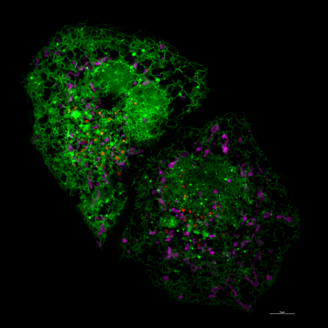
A super resolution image captured in Diao's lab shows the endoplasmic reticulum in green, lysosomes in pink and mitochondria in red. Photo provided by Jiajie Diao.
In the first use of the new measurement technique described in the article, the researchers found that mitochondria, the organelle in cells responsible for energy production and respiration, become enlarged when developing into degenerative diseases.
Lysosomes were also found to tend to move closer to mitochondria when the mitochondria are damaged. Further research can help determine more about how organelles’ sizes and placements in relation to each other can cause disease, Diao said.
Increasing capabilities with AI
Now that the measurement imaging technique has been developed, Diao said the team is beginning to work with computer scientists to harness the power of artificial intelligence to increase how it can be used.
“Nowadays, lots of measurements are made by people and by a simple algorithm, so we still need manpower,” Diao said. “We are developing AI, the machinery, the intelligence to try to do everything by the machine.”
Down the road, Diao said the hope is that an algorithm can be trained with a wide variety of images of healthy and diseased cells so that the software can analyze a cell image and then predict whether or not it will become cancerous.
The technique combined with AI could also be used to study the effectiveness of drugs to treat diseases, for example to see if there are specific organelles that are contributing to a patient developing drug resistance to a certain medication.
“This will give us a next level ability,” he said. “Currently, most people are looking at the tissue level or a higher level, but we can go down to the subcellular level.”
Due to the purchase of a new microscope from the UC College of Medicine and Department of Cancer Biology, any researcher at the University of Cincinnati or Cincinnati Children’s Hospital Medical Center is now equipped to take advantage of the innovations from Diao’s lab in future studies.
“So that’s what we actually are trying, to let more people use the most advanced probe and the most advanced imaging technique to do their study,” Diao said.
Young Investigator Award

Jiajie Diao, PhD. Photo/University of Cincinnati.
Diao was recently named the recipient of the 2022 Young Investigator Award by the UC Chapter of Sigma Xi. The annual award, presented in conjunction with the UC Office of the Vice President for Research, recognizes a junior faculty member at UC for their early career distinguished research accomplished in a field of science or engineering appropriate to Sigma Xi.
Jun-Lin Guan, PhD, the Francis Brunning professor and chair of the Department of Cancer Biology, said the award is well-deserved.
Diao was the first junior faculty recruited to the department after Guan assumed the chair position in 2014. Since joining the department in 2015, Guan said Diao has made a number of advancements in the monitoring and study of subcellular dynamics of organelles and proteins in several cellular processes and has published more than 50 manuscripts in various journals.
Guan said Diao’s expertise in physics made him an atypical candidate for a position in a cancer biology department, but his “passion in science, superior training and publication track record in interdisciplinary research” has helped him become a quality faculty member.
“I would say it was one of the best early decisions that we made back then, and now we are seeing fruits several years later,” Guan said.
Diao’s continued success bodes well for the future of basic science and cancer biology research at UC, Guan said.
“This is top-notch basic science research but with significant relevance for studies of cancer cells and signaling, the major focus area for the University of Cincinnati Cancer Center basic science program,” he said. “[Diao] serves as a model for our aspiring young junior scientists in the department and at UC. Together, there are greater things to be achieved for basic science research and cancer programs at UC.”
Diao will be presenting highlights of his research at the Sigma Xi Spring Mixer on March 8 at the Mantei Center, the engineering research center on the Uptown campus. A social mixer will begin at 4:30 p.m. outside of room 427, with the formal program and presentation beginning around 5:15 p.m. inside room 427.
Featured photo at top of super resolution image of cell courtesy of Jiajie Diao.
Next Lives Here
The University of Cincinnati is classified as a Research 1 institution by the Carnegie Commission and is ranked in the National Science Foundation's Top-35 public research universities. UC's medical, graduate and undergraduate students and faculty investigate problems and innovate solutions with real-world impact. Next Lives Here.
Related Stories
How scammers can persuade you with AI
July 27, 2026
Spectrum News recently dove into how cloning voices with artificial intelligence is making it easier to scam people over the phone. The news outlet spoke with Kimberly Hyun, assistant professor of marketing at UC’s Lindner College of Business, to examine why AI is making it so much easier to trick people.
Decade of discovery
July 27, 2026
Every summer, UHP Discover has connected undergraduate students in the University Honors Program (UHP) to a unique array of opportunities in original interdisciplinary research. This year, the program celebrates its 10th anniversary.
Building community
July 27, 2026
The inaugural Ultra-SLP Conference brought together clinicians and researchers from across the country to advance the practice of using ultrasound technology to treat disorders of speech and swallowing.