New probe aids novel findings on cell functions
Collaborative team publishes research in ACS Sensors

Collaborative research at the University of Cincinnati has developed a new probe to better study cells that has already led to new knowledge about certain cellular processes.
UC’s Jiajie Diao, PhD, and Yujie Sun, PhD, are lead authors on new research published May 4 in ACS Sensors.
Focus on endolysosomes
The team’s research focused on organelles, or specialized structures that perform various jobs inside cells, called endolysosomes. Lysosomes are organelles that act as the “recycling center” of the cell, reusing broken or malfunctioning building blocks for different purposes, and endolysosomes are a subset of lysosomes that begin as a different organelle called an endosome.
Lysosomes are an important organelle to study because abnormalities can lead to what are called lysosomal storage diseases that cause buildups of toxic substances in cells. Abnormalities in lysosome function are also associated with neurodegenerative diseases and cancer.
“If lysosomes are not functioning normally, then the cell will accumulate lots of waste, eventually leading to cell death,” said Diao, University of Cincinnati Cancer Center member and associate professor in the Department of Cancer Biology in UC’s College of Medicine.
In the process of transitioning from an endosome to a lysosome, these organelles change their pH levels from a neutral environment to an acidic environment. If the environment is not acidic enough, lysosomes cannot do their jobs of clearing and recycling waste properly.
“We are trying to image this process and trying to track the whole changing process of the endolysosome and trying to figure out in which state and which location these endolysosomes will become abnormal,” Diao said.
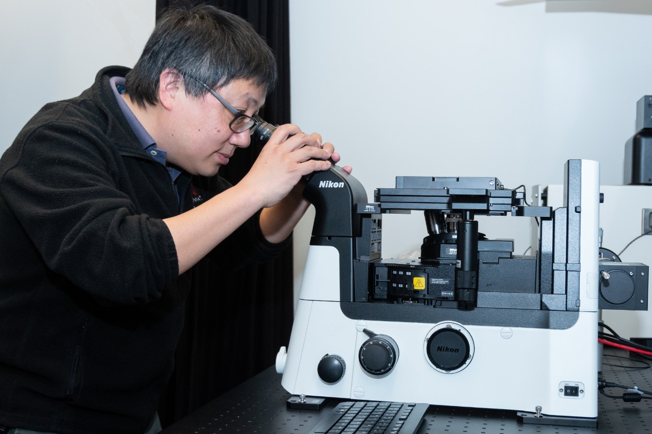
Diao works in his laboratory in the Vontz Center. Photo/University of Cincinnati Cancer Center.
New probe
Diao and Sun’s team published research last year on the ECGreen probe that turned a brighter shade of green when the cell environment becomes more acidic. While it was a step forward, Diao said ECGreen still had limitations.
“It’s hard to gauge the absolute value, because I can only know it’s become lower,” Diao said. “The intensity becomes higher, but how high is high? There’s no way you can actually calibrate this value.”
The newest probe the team developed is green when in a neutral environment and shifts to a red color when the cell environment turns more acidic.
“So we have red signaling and green signaling, and by calculating the ratio, we can calibrate the exact pH value,” Diao said. “This is more robust and more precise to measure the pH inside the endolysosome, so this is a big advantage.”
“Our probe exhibits excellent pH-sensitive fluorescence in endolysosomes at different stages of interest,” added Sun, a Cancer Center member, associate professor and graduate program co-director in UC’s Department of Chemistry.
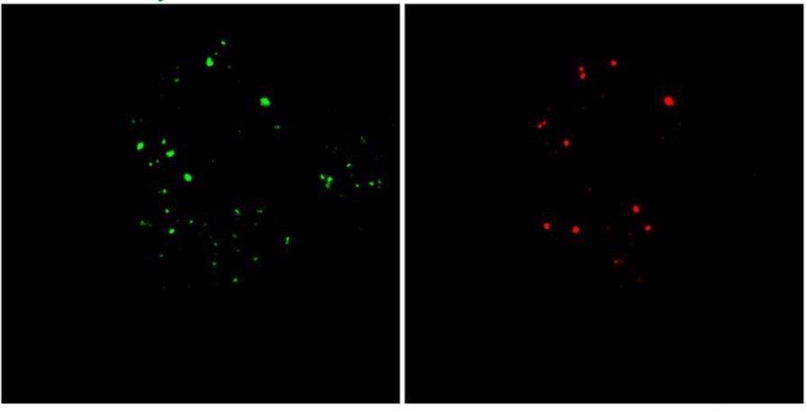
A HeLa cell labeled by the new probe shows total endolysosomes in green, with late endosomes/lysosomes in red. Photo provided by Jiajie Diao.
Research in action
When a cell’s environment changes, like when a cell is damaged, it needs to increase the number of lysosomes in order to recycle or clean up the cell. But the process cells use to increase the number of lysosomes was not previously known.
Using the new probe, the researchers found no matter what environment a cell is in, and whether it is a normal or abnormal cell, it will keep a constant ratio of endosomes that it then converts into lysosomes.
“By applying the small molecular probe in live cells, we were able to reveal a constant conversion rate from early endosomes to late endosomes/lysosomes,” Sun said. “We feel quite happy we were able to reveal this constant conversion rate.”
Diao noted that for the first time, the team found this process from endosome to lysosome is governed by a complicated protein-controlled process.
“This process is highly regulated, and you cannot skip any steps, even if it’s an emergency,” Diao said.
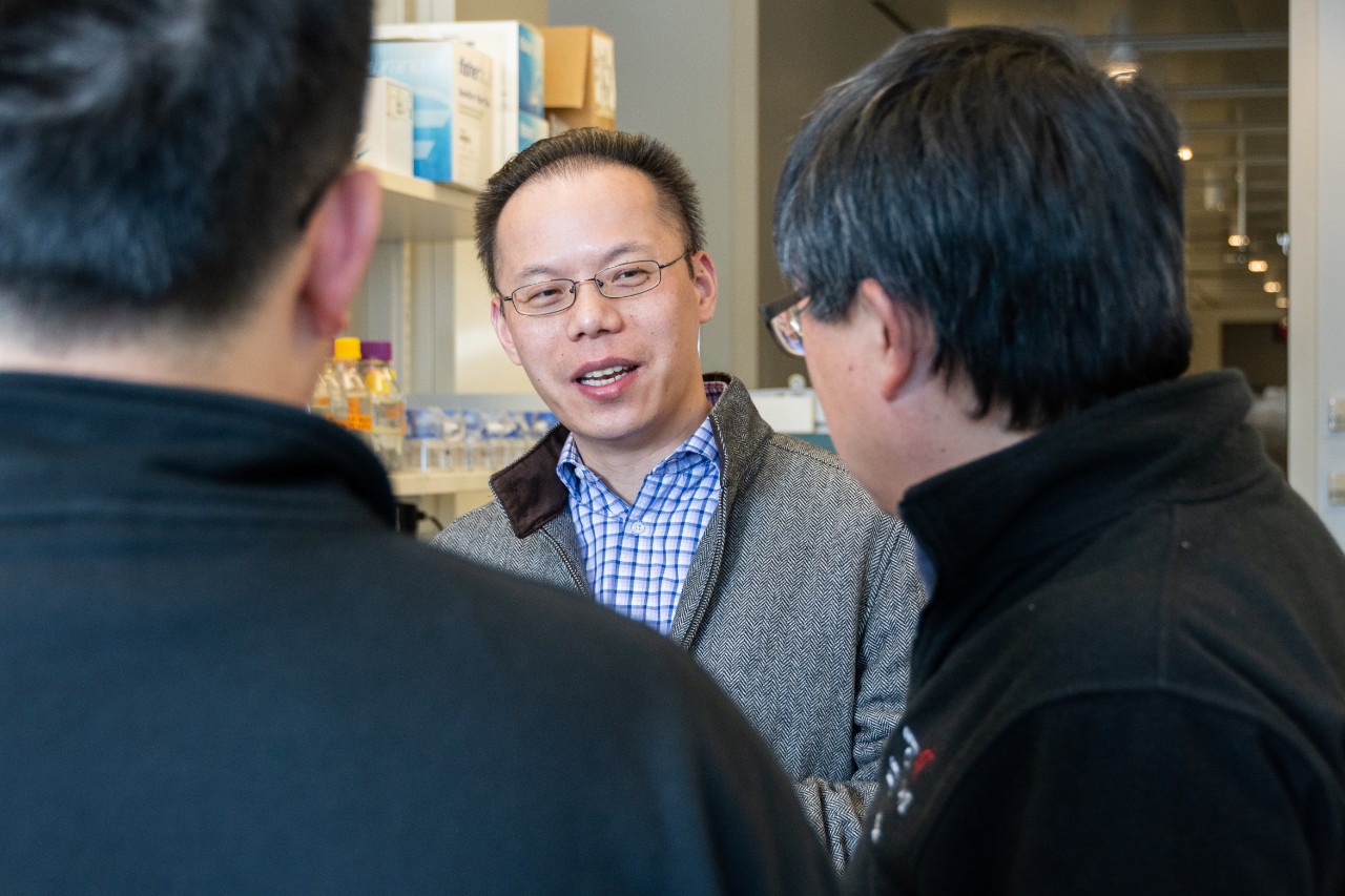
Sun, center, Diao, right, and their teams have become close collaborators to move research forward. Photo/University of Cincinnati Cancer Center.
Probe applications
In addition to the uses described in the published research, Diao said there are a number of other applications for the new probe.
“We can know the location of the endolysosome, we can know the number, we can know the pH change, we can know the size of the endolysosome,” he said. “Eventually we will have all parameters about the endolysosomes inside the cell.”
With all of this information, the team is working to develop a machine-learning software that will be able to profile cell abnormalities and quickly diagnose lysosome problems that may lead to diseases. This method would be faster and more cost effective than current gene and protein sequencing, and in practice could only be a healthy patient taking a simple blood test that tells them if they have risk factors for certain lysosomal diseases.
“You just need to do staining, and in 10 minutes, you have all the information,” Diao said. “We’re actually trying to develop a system with these probes so we can do a fast diagnosis.”
The researchers are also studying treatments that affect the process of transition from endosome to lysosome to speed up the complicated process, including research using light to speed up the process.
“Our research results demonstrate that it is an extremely promising direction in designing and developing small fluorescent probes for bioimaging applications, especially when coupled with high-resolution microscopy,” Sun said. “We will continue developing novel fluorescent probes with multiple functions.”
Other researchers at UC have begun to use the probe, and the team is working to patent and commercialize the technology.
“I think this is a really classic work resulting from collaboration,” Diao said. “Dr. Sun and our group work together and we actually make the whole process very fast and very efficient. We want to accelerate all this discovery.”
“Complementary expertise of Dr. Diao's and my own groups really make it possible to work on these collaborative projects,” Sun added. “The synergistic interaction between our groups is the key of success.”
Next Lives Here
The University of Cincinnati is classified as a Research 1 institution by the Carnegie Commission and is ranked in the National Science Foundation's Top-35 public research universities. UC's medical, graduate and undergraduate students and faculty investigate problems and innovate solutions with real-world impact. Next Lives Here.
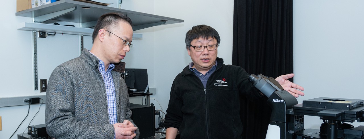
Featured photo at top of Sun, left, and Diao, right in the laboratory. Photo/University of Cincinnati Cancer Center.
Related Stories
Three years, countless stories
May 15, 2026
UC's Klekamp Law celebrates its 193rd Hooding with stories from graduates reflecting on their paths through the college.
Driven by curiosity, guided by care
May 14, 2026
Max Wilson, a University of Cincinnati College of Allied Health Sciences health sciences major on the pre-physician assistant track, found his path expanding beyond the classroom and into hands-on research focused on human performance and patient care.
UC Blue Ash celebrates top students and recognizes Honor Student of the Year
May 14, 2026
The University of Cincinnati Blue Ash College recently hosted a special event that celebrated students for exceptional achievements during the 2025-26 academic year. The honorees included academic award winners, student engagement award winners, Latin Honors graduates, and the 2026 UC Blue Ash College Honor Student of the Year.